-
08:00 - 17:00


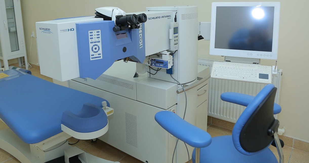
Laser correction
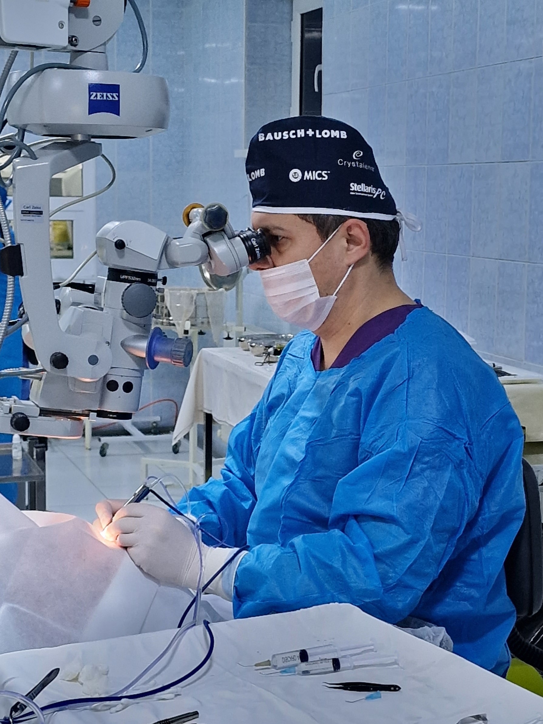
Surgical treatment
The highest difficulty
1. IOL implantation by scleral fixation
with anterior vitreoectomy
2. Removal of dislocated lens + IOL (p/c)
3. Anti-glaucamatous surgery on the only eye
Survey
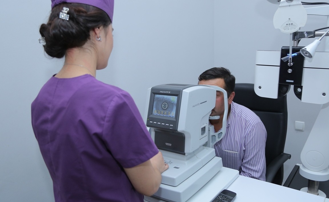
Diagnostic examination:
1. Determination of visual acuity without and with correction.
2. Secondary examination, Keratometry.
3. Goldmon tonometry.
4. Determination of the nature of vision.
5. Biomicroscopy.
6. Ophthalmoscopy.
7. Consultation of an ophthalmologist.
8. Ultrasound of the eyeball.
Additional diagnostic examination:
1. Tomography.
2. Computer echobiometry.
3. OCT.
4. Kenalog (introduction to the subtenolic space of the eyeball).
5. Kenalog (intravenous administration of drugs).
6. Laser coagulation of the retina.
7. Fluorescence angiography (FAG).
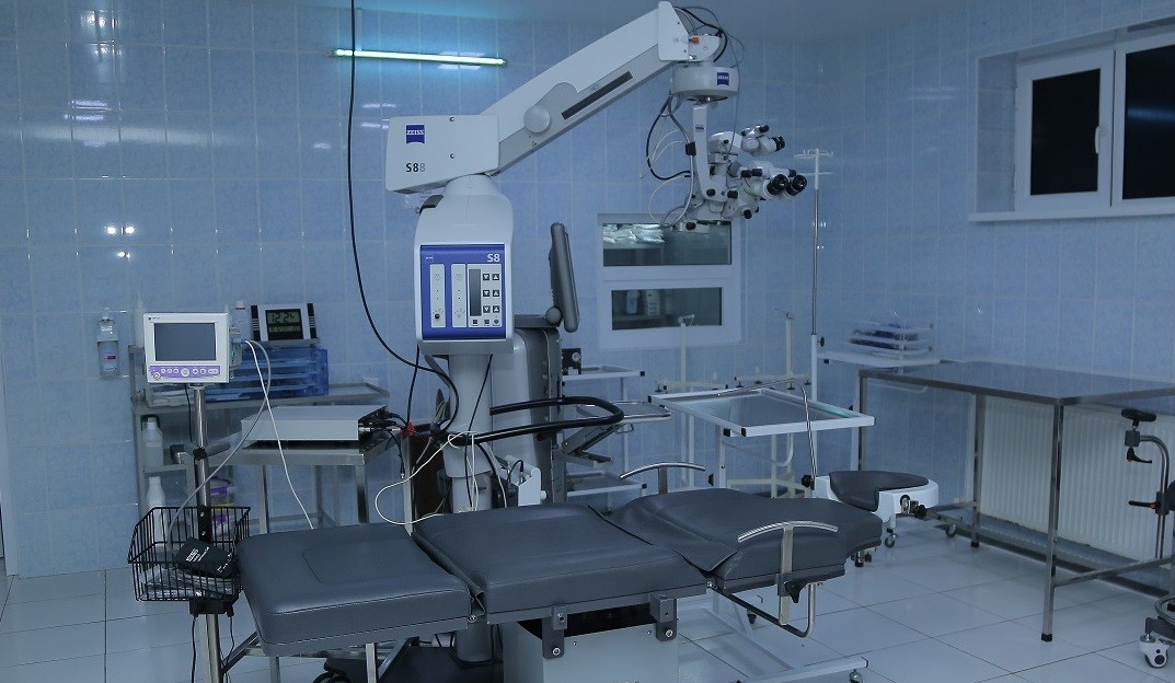
Vitreoretinal Surgery
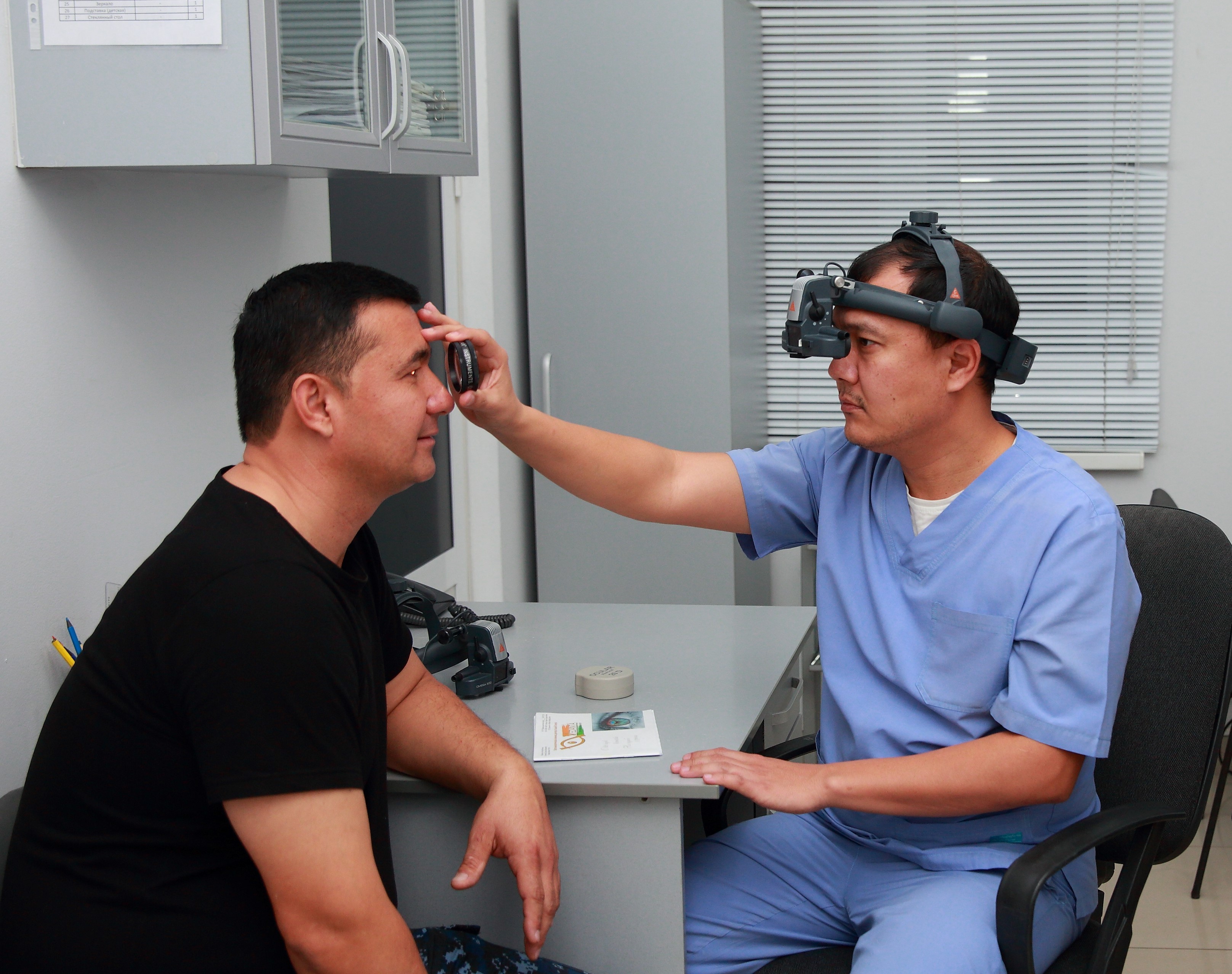
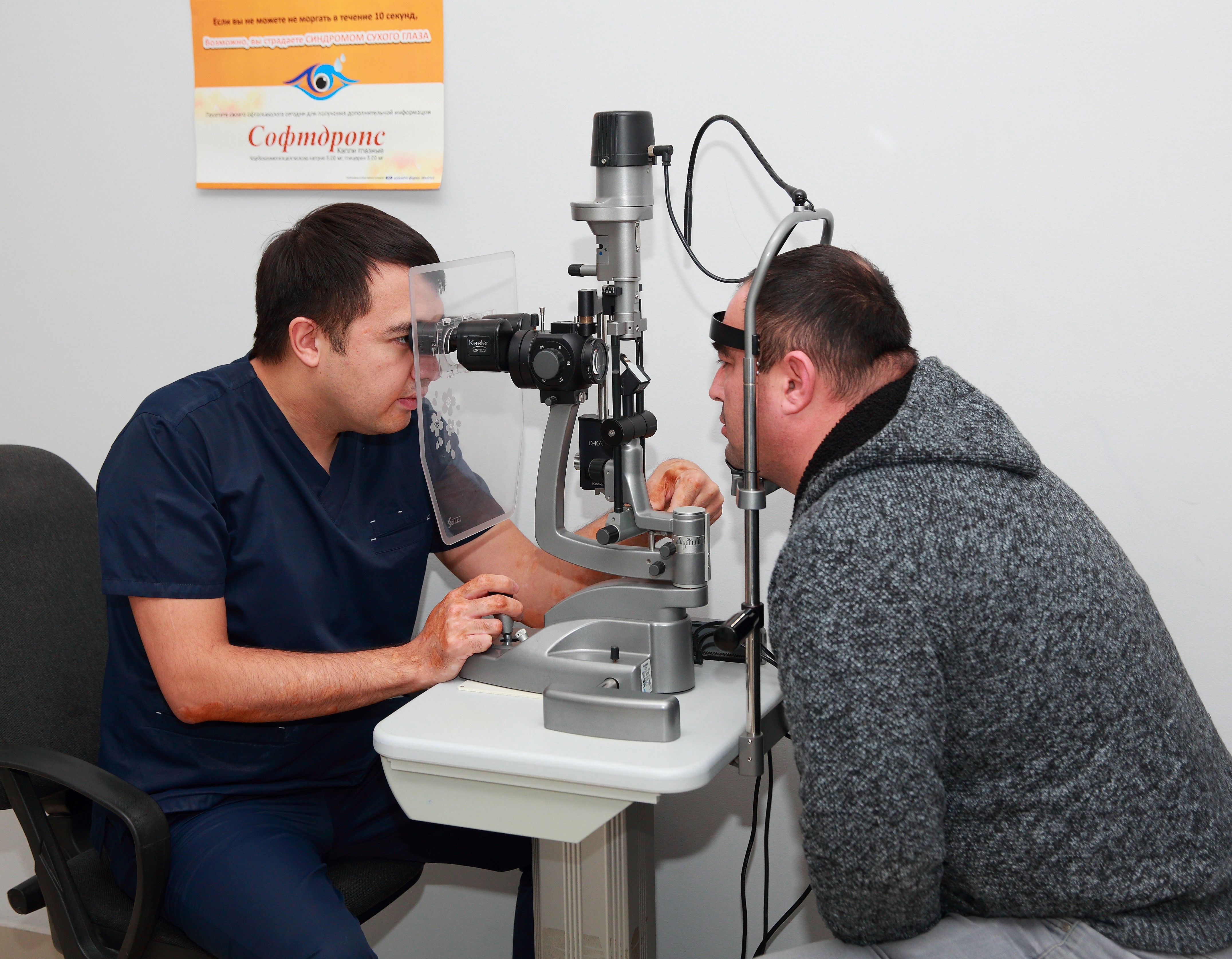
Ophthalmologist consultation
The eyes provide a person with more than 85% of all information. Visual impairment, and even more so its loss, leads to a limitation of a person's natural abilities and discomfort, significantly reduces the quality of human life. Therefore, timely consultation with an ophthalmologist is important, which can help avoid serious consequences. An ophthalmologist is a doctor who is engaged in the diagnosis and treatment (therapeutic, optical, cosmetic and surgical) of diseases of the eyes and auxiliary apparatus (eyelids and lacrimal glands).
When should I consult an ophthalmologist? Quite often, eye diseases are asymptomatic. Therefore, in order to detect them in a timely manner, it is required to visit an ophthalmologist annually. The child must be brought to consult a specialist twice a year. Pregnant women and people suffering from diabetes mellitus, atherosclerosis, diseases of the cardiovascular system, liver and kidney diseases, impaired functioning of the endocrine system should regularly undergo vision diagnostics. An extraordinary visit to the doctor will be needed for: pain, burning, itching in the eyeball; visual defects; photophobia; increased lacrimation; bleeding; changes in the field of vision; decreased visual acuity; distortion of the shape of objects; the appearance of circles, dots, "midges", fog in front of the eyes; redness of the eyelids or eyeball; white pupil; dry eyes; swelling and inflammation of the eyelids; trichiasis – the wrong direction of eyelash growth; purulent discharge from the eyes; the appearance of a yellow plaque on the shell of the eye; the formation of barley. Diagnosis The success of treatment depends on the correct diagnosis. At the initial appointment, the doctor first examines the patient's complaints and anamnesis of the disease.Next, studies are assigned that can help in the diagnosis.